One person arrives with pain in the temple and is certain it is a migraine. Another has an aching arm and a referral for neck imaging. A third limps every morning because of a “heel spur”. The pain is real in all three cases, but the place where it speaks does not match the place where a muscle is overloaded. This is neither a mystery nor esoterics: referred pain has a rather dull explanation in the wiring of the nervous system, and a practical map that you can follow with your hands.
What a trigger point is, and what is still being argued about
The working definition is simple. A trigger point is a small tender spot inside a taut band of muscle fibres which, under pressure, produces not only local pain but a recognizable pain somewhere further away. The key word is recognizable: on the table people usually do not say “that hurts”, they say “yes, that is the pain, that is the one I live with”.
Points are divided into active and latent. An active one hurts on its own, with no pressure at all, and that is the one people come in with. A latent one stays quiet until a finger finds it, yet it already shortens the muscle and limits range — which is why someone who reports that “nothing hurts” cannot comfortably turn the head all the way to the shoulder or sit back on their heels.
Now for the honest caveat, without which there is little point reading on. A trigger point is a clinical model, not a proven anatomical structure. There is still no reliable image that would show one on ultrasound or MRI. In studies, agreement between different practitioners palpating the same back is low: what I would call a taut band, a colleague may not find. The popular explanation involving excess acetylcholine release at the motor endplate, local ischaemia and sensitization of nociceptors is a hypothesis, and it has both supporters and serious critics.
What is barely disputed is the phenomenon of referred pain itself. Pressure at one spot reproducibly creates pain elsewhere, and the routes repeat from person to person so consistently that they have been drawn on charts for more than half a century. The argument is about what exactly we are feeling under the fingers and why manual work helps. It is not about whether a predictable map exists.
A trigger point is not a diagnosis. It is an address worth starting from when the pain speaks somewhere other than where it would make sense.
Ale
So in the studio I rely on signs that can be checked right now, rather than on an elegant theory:
a dense band can be felt under the finger, running along the fibres rather than across them;
somewhere in that band is a point far more tender than the centimetres either side of it;
pressure reproduces the exact pain the person came in with;
the pain travels in a predictable direction instead of spreading at random;
the muscle is shortened: range on that side is smaller, and the difference is visible to the naked eye;
occasionally a cross-fibre stroke makes the fibres twitch briefly — a local twitch response.
Four classic routes that explain strange pain
Muscles in focus
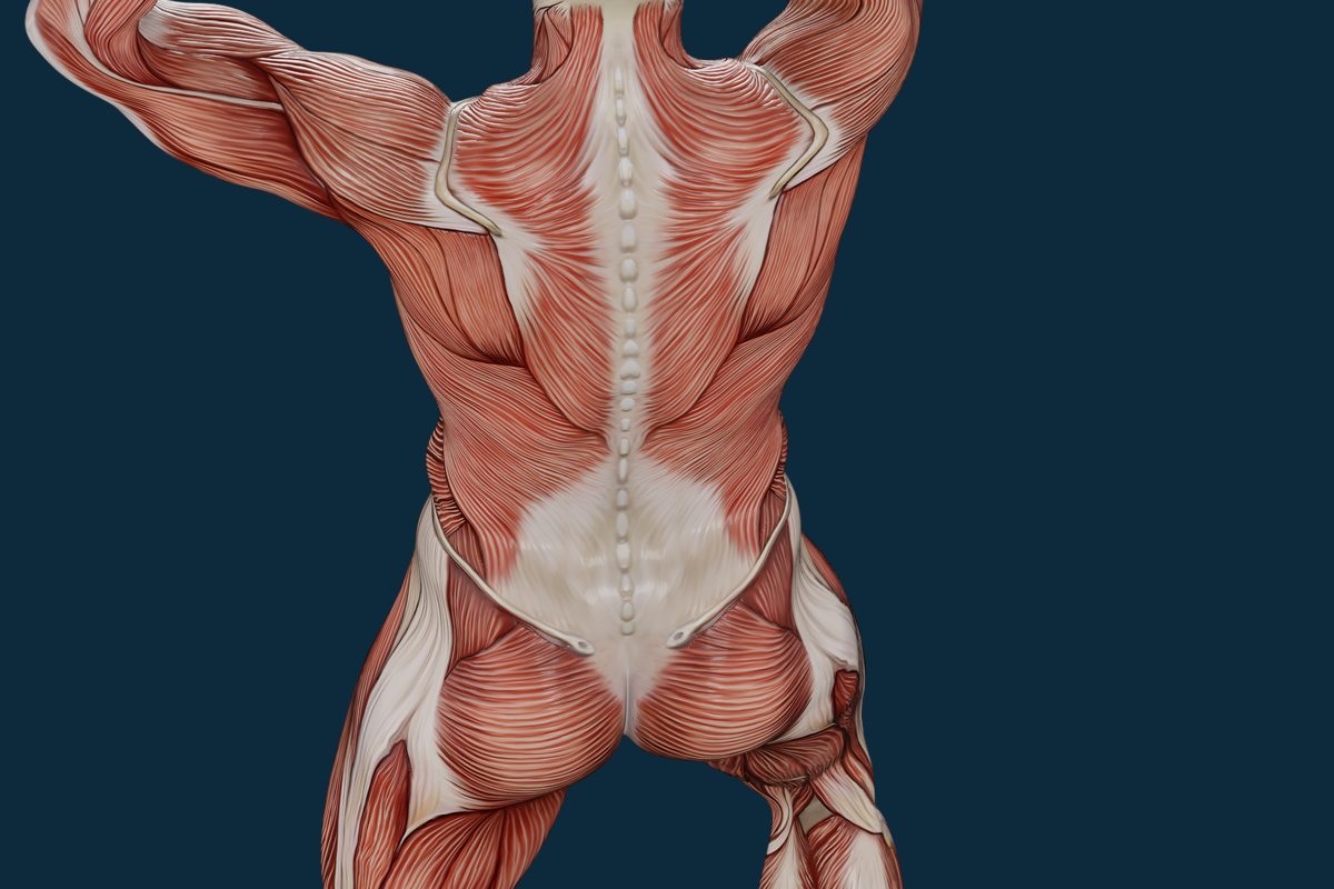
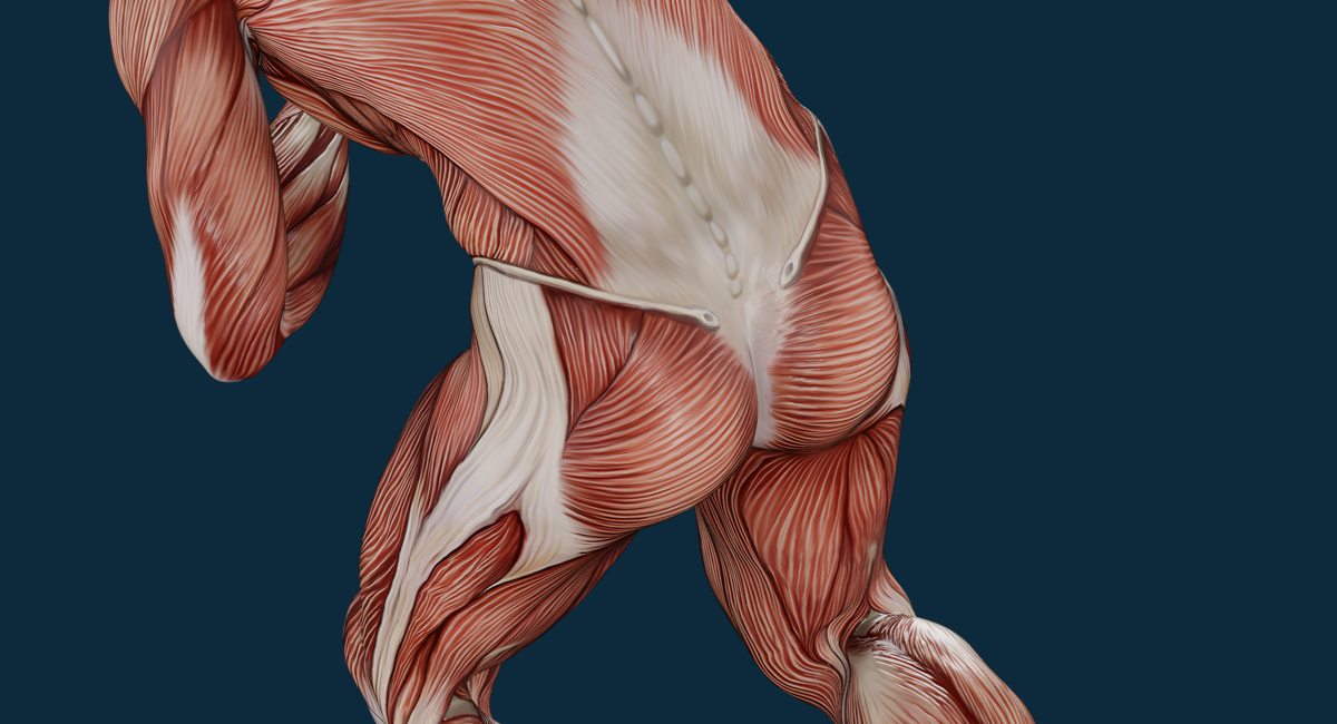
Rear view, arms raised: the trapezius (m. trapezius) in full, the deltoids, latissimus dorsi, the erector spinae (m. erector spinae), the thoracolumbar fascia, the glutes, the hamstrings and the calves. Half the referral routes in this article begin on this frame.
Trapezius (m. trapezius)
Deltoid (m. deltoideus)
Latissimus dorsi (m. latissimus dorsi)
Erector spinae (m. erector spinae)
Thoracolumbar fascia (fascia thoracolumbalis)
Glutes (mm. glutei)
Referral map
Four routes on a body map
Tap a point to light up the area it usually refers to.
Front
Back
1trigger point — tap it
main referral zone
spillover zone — not in everyone
Points
1Point · Back
Upper trapezius
m. trapezius, pars descendens
Where the point is
Middle of the shoulder ridge, between the neck and the shoulder.
Where it refers
Back and side of the neck, behind the ear, temple; sometimes the angle of the jaw.
What usually sets it off
Screen below eye level, phone held by the shoulder, bag on one side.
Zone shown: Front · Back
If the “migraine” came on suddenly and is the worst of your life, or comes with slurred speech, weakness or numbness on one side of the body, it is an emergency.
2Point · Front
Scalene muscles
mm. scaleni
Where the point is
Side of the neck, above the collarbone.
Where it refers
Chest, shoulder, outer side of the arm, forearm, thumb and index finger; the inner edge of the shoulder blade.
What usually sets it off
Upper-chest breathing, stress, coughing, carrying heavy loads, driving.
Zone shown: Front · Back
Numbness and weakness in the hand, or numb fingers at night — see a neurologist.
3Point · Back
Gluteus minimus
m. gluteus minimus
Where the point is
Deep under the gluteus medius, in the outer part of the buttock.
Where it refers
Outer and back of the thigh and lower leg, sometimes to the ankle — “like sciatica”.
What usually sets it off
Long sitting, sleeping on that side, running on cambered ground.
Zone shown: Front · Back
Numbness, a weak foot, pain in a narrow strip, worse with coughing — signs of a nerve root, see a neurologist.
4Point · Back
Soleus
m. soleus
Where the point is
Lower part of the back of the leg, under the gastrocnemius.
Where it refers
Back and underside of the heel, less often the sacral area.
What usually sets it off
New shoes, hard surfaces, a sharp jump in running volume.
Zone shown: Back
A swollen calf that is red, warm and firm may be a thrombosis: massage is contraindicated, see a doctor at once.
The map is a working model, not a diagnosis. Numbness, weakness, shooting pain — see a doctor, not a massage table.
Patterns after Travell & Simons, “Myofascial Pain and Dysfunction: The Trigger Point Manual”. Each zone is drawn on one side of the body for clarity, although it can occur on both; in a given person the zone may be smaller or shifted. A diagram, not a measurement.
Upper trapezius to the temple. The most frequent route in my practice. The pain climbs the side of the neck, passes behind the ear and settles in the temple, sometimes in the angle of the jaw or behind the eye. People describe it as “a migraine that starts after lunch and becomes unbearable by the evening”. The context is almost always ordinary life: a laptop sitting below eye level, a phone pinned by the shoulder during calls, a bag carried on the same side for ten years. The muscle is neither torn nor inflamed — it simply holds the head in a slightly awkward position for eight hours a day.
Scalenes to the arm. The scalenes (mm. scaleni) lie on the side of the neck and lift the top two ribs: in quiet breathing their contribution is small. But if you breathe into the upper chest — under stress, after a long bout of coughing, or from a habit of holding the stomach in — they take over as primary breathing muscles and become overloaded. The referral runs into the shoulder, down the outer side of the arm, into the forearm, often into the thumb and index finger, and sometimes under the shoulder blade and forward into the chest. It reads as “a pinched nerve”, and people quite reasonably expect a scan of the neck.
Gluteus minimus to a leg that feels like sciatica. Gluteus minimus (m. gluteus minimus) is deep, lying beneath gluteus medius, and it is not visible from the outside on any frame. Its referral travels down the outer or back of the thigh to the lower leg, sometimes almost to the ankle. This is the classic imitation of radicular pain: the person has already said the word “sciatic”, is already perching on the edge of the chair and is already afraid to bend forward. The difference is in the details — more on those below.
Soleus to the heel. Soleus (m. soleus) lies deeper than gastrocnemius (m. gastrocnemius) and works when the knee is bent: on a climb, on stairs, on every step over hard ground. Its points refer into the back and underside of the heel and, rarely, into the area of the sacrum — the most unexpected route on the whole map. Morning pain, that first step like a nail going in, is usually blamed on a spur, even though the bony outgrowth turns up on the scans of people with no complaints at all. Stubborn heel pain belongs with a doctor, but the lower leg is worth checking either way.
Muscles in focus
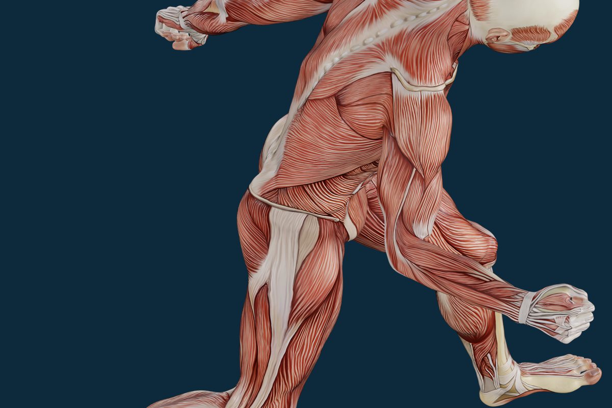
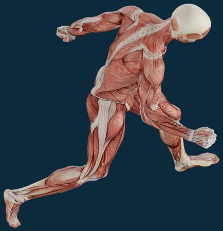
Running stride, rear three-quarter view: the glutes, the hamstrings, the calf (m. gastrocnemius) and the Achilles tendon. Soleus lies underneath the calf and is not visible in the frame — which is precisely why it is so rarely suspected in pain under the heel.
Glutes (mm. glutei)
Hamstrings (mm. ischiocrurales)
Gastrocnemius (m. gastrocnemius)
Achilles tendon (tendo calcaneus)
Where the point is
Where it refers
How the complaint sounds
Typical context
Upper trapezius
Side of the neck, behind the ear, temple, angle of the jaw
“A migraine starts up by the evening”
Screen below eye level, phone held by the shoulder, bag on one side
Scalenes
Shoulder, outer side of the arm, forearm, thumb and index finger
“It feels like a pinched nerve in my neck”
Upper-chest breathing, stress, coughing, carrying heavy loads, driving
Gluteus minimus
Outer and back of the thigh and lower leg, sometimes to the ankle
“Sciatica, it shoots down my leg”
Long sitting, sleeping on that side, running on cambered ground
Soleus
Back and underside of the heel, less often the sacral area
“A spur: the first step in the morning is like a nail”
New shoes, hard surfaces, a sharp jump in running volume
Why it hurts away from the cause
The mechanism is called convergence. Fibres from skin, muscles, joints and internal organs arrive at the same segment of the dorsal horn of the spinal cord. From there a shared wire runs upward, and the brain, receiving the signal, has to decide where it came from. With skin there is no difficulty: its representation in the cortex is large and we train that precision all our lives — catch a corner of a table and you can point to the spot within a centimetre. Deep tissue is another matter: muscular pain is dull, diffuse, “somewhere around here”, and the brain regularly gets the address wrong, assigning it to a neighbouring territory of the same segment.
The second mechanism is sensitization. When a stream of pain signals continues for weeks, dorsal horn neurons lower their threshold and widen their receptive fields. In practice it looks like this: at first a coin-sized spot hurt, a month later half the shoulder blade aches, and a month after that “something similar” shows up on the other side, where the tissue is perfectly healthy. This is not damage spreading through the body — it is the gain on the amplifier being turned up.
The best known example of convergence has nothing to do with muscles at all: in a heart attack the heart is the organ under strain, yet the pain speaks in the left arm and the jaw. Nobody blames the arm. With muscles the principle is the same, only the cost of a wrong address is usually lower — someone spends six months treating a temple instead of raising the monitor.
There is one practical conclusion: the map is read backwards. Pain in the temple — look first at the neck and the upper trapezius. Shooting into the arm — check the scalenes and pectoralis minor (m. pectoralis minor). “Sciatica” with no neurological signs — go to the glutes. A heel in the morning — look at the lower leg. This is not dogma, just the order in which the hypotheses are cheapest to test by hand.
Ischaemic compression: what the hands do and what you feel
First the search. I work across the fibres with the pads of my fingers, find the band and follow it to the point where tenderness is sharply higher than in the centimetres around it. Then pressure: with a finger, a knuckle or an elbow if the muscle is large and lies deep. It builds gradually, over several seconds, rather than dropping in all at once.
Then holding. I hold the pressure until the pain under the finger drops by roughly half — usually anywhere from twenty seconds to a minute and a half. If nothing changes in that time, I do not press harder, I let go: it means the hypothesis was wrong or the tissue is not ready today. After the compression comes a slow glide along the band, and then movement, without exception — active movement, made by your own effort. The muscle needs the experience of working at its new length, otherwise it will return to the familiar one within an hour.
Now about dosage, because this is the great misunderstanding around deep work. The working range is 6-7 out of 10: clearly felt, definitely unpleasant, but you stay in contact with what is happening. The simple criterion is your breathing. If you can breathe, the pressure is right. If you hold your breath, press yourself into the table, or your shoulders start creeping up, the body is answering with defence rather than release, and I will spend the next twenty minutes undoing the effects of my own pressure. Deep tissue does not mean “put up with it if it hurts”, and bruises are neither the goal nor a sign of good work.
Muscles in focus
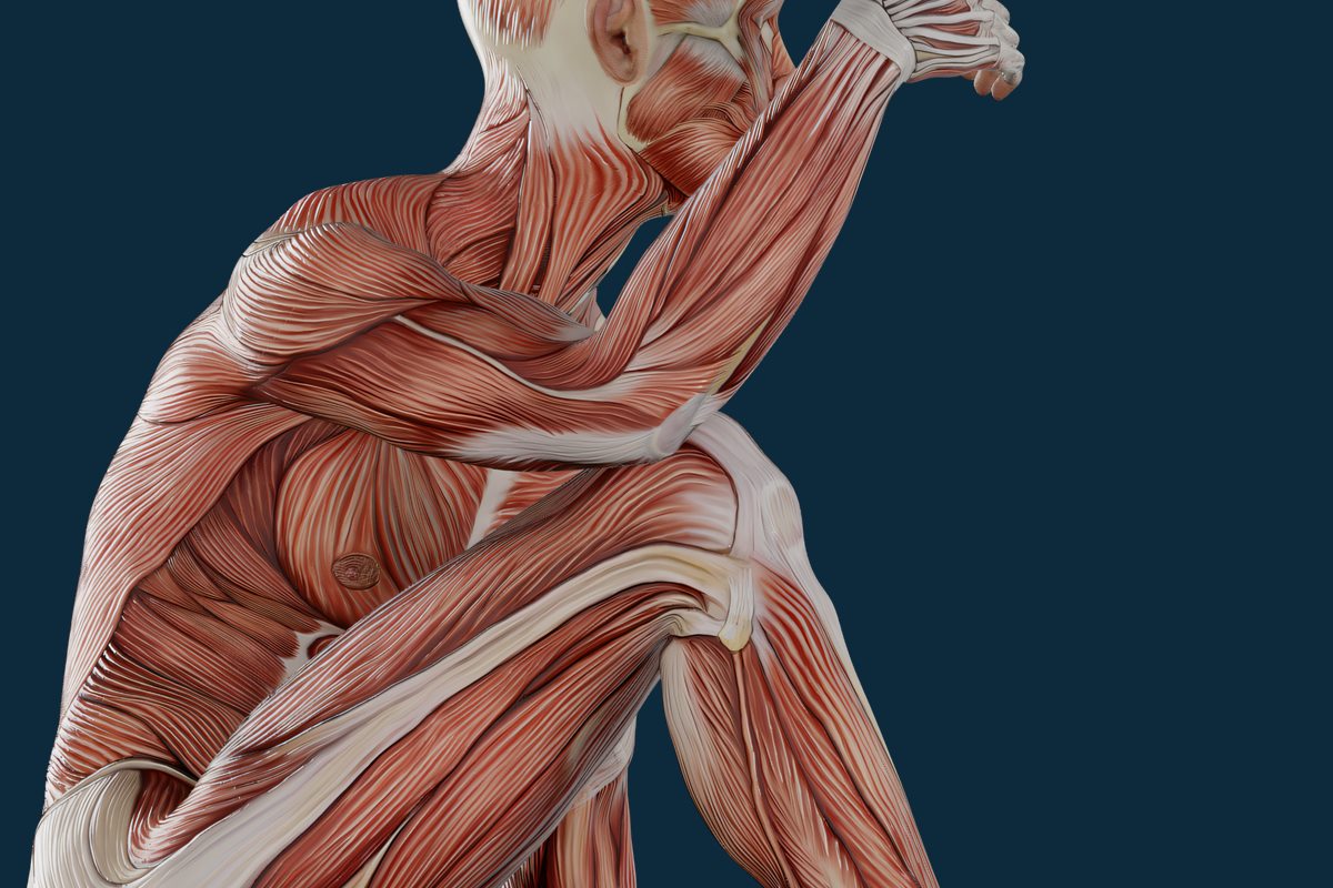
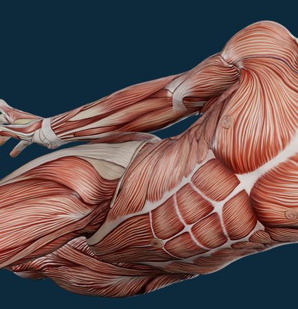
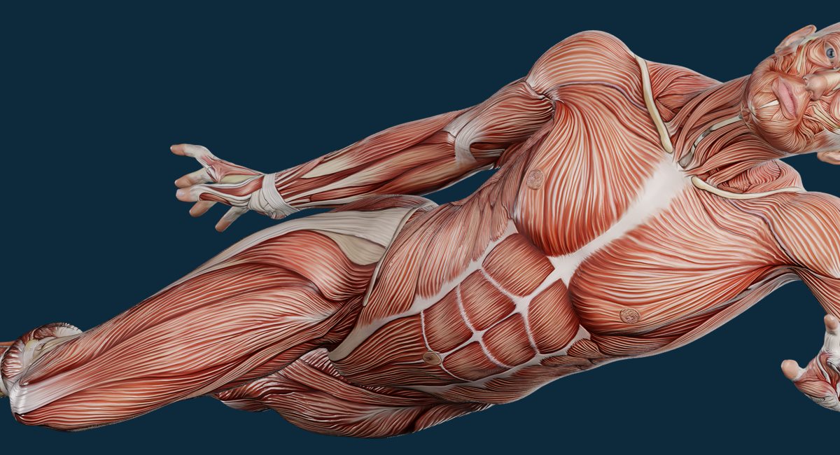
Seated, arms around the knees: the neck and the upper trapezius, the deltoid, the erector spinae, the glutes, the calf. This is the position in which the upper trapezius is at its most shortened — and the one from which it most often refers into the temple.
Upper trapezius (m. trapezius, pars descendens)
Deltoid (m. deltoideus)
Erector spinae (m. erector spinae)
Glutes (mm. glutei)
Gastrocnemius (m. gastrocnemius)
What you feel, step by step: first recognition — “that is the pain”. Then referral: the sensation travels along its route, and many people see the link between neck and temple for the first time. Then the pain under the finger goes dull, spreads out and fades — that is the signal to release. After a session most people feel warmth and heaviness in the area, sometimes drowsiness, and over the next 12-48 hours there may be soreness rather like the day after an unfamiliar workout: that is normal and passes on its own.
And about the evidence for the technique — as plainly as about the model itself. Reviews show that manual work on trigger points reduces pain and improves range in the short term, but the quality of the studies is middling on average, and no advantage has been proven for ischaemic compression specifically over other forms of manual work. Put simply: the effect is real, useful and noticeable, but it does not rebuild your posture and it does not cancel the load that keeps recreating the point. If nothing changes in how you sit, how you carry your bag and how you add running kilometres, the point will quietly come back within two to six weeks.
When it is not a trigger point but a reason to see a doctor first
Referral can usually be told apart from radicular pain by simple signs. When a nerve root is compressed, the pain runs in a narrow, well defined stripe, joined by numbness, tingling or weakness; reflexes are reduced, and coughing or straining clearly makes the symptom worse. Referred muscular pain is dull, its area is blurred, sensation and strength are intact, but pressing one specific point reproduces exactly that pain. This is not a test that replaces a neurological examination, but it is a difference you can notice yourself and describe clearly to a doctor.
Red flags: the doctor first, hands afterwards
Weakness, numbness or a dropping foot or hand. Numbness in the groin area, or changes in bladder or bowel control. Fever together with back pain. Night pain that does not depend on position, especially with weight loss. A history of cancer. A recent injury, fall or road accident. Chest, jaw or left arm pain on exertion, with shortness of breath or a cold sweat, is an emergency: call an ambulance, not a massage therapist. Pain and swelling in the calf with redness, warmth and firmness may be a thrombosis, and massage is categorically contraindicated. A first-ever, worst-ever headache, or a headache with fever and a stiff neck. In all of these cases a session is not merely useless — it steals time.
Let me restate the general limits separately, because they apply to trigger point work too: fever and respiratory infections, acute inflammation, thrombosis, thrombophlebitis and varicose veins in the working area, cancer — only with the agreement of your treating doctor, recent injuries and fractures, skin infections in the working area, uncontrolled high blood pressure, anticoagulant therapy, pregnancy — which calls for a specialist in that field, and intoxication. The full list with explanations is gathered in the answers to frequent questions.
And the main thing. Massage is not medical care: I do not make diagnoses and I do not prescribe anything. My job is to test the muscular explanation by hand and tell you honestly whether it holds up. If the picture has not changed after two sessions, or any of the symptoms above appear, the right next step is a doctor, not a third session. How the work itself is structured is described in detail on the page about deep tissue massage, and you can look at the regions and muscles in the atlas.
Key points
A trigger point is a working clinical model, not a proven structure: the referral map is reproducible, the mechanism is still under discussion.
Four common routes: upper trapezius to the temple, scalenes to the arm, gluteus minimus to the leg, soleus to the heel.
Referral is explained by convergence of signals in the dorsal horn and by sensitization — nervous system mechanics, not metaphors about “knots”.
Ischaemic compression works in the 6-7 out of 10 range: if you can breathe, the pressure is right, and bruises are not the goal.
Numbness, weakness, fever, night pain, a swollen calf or chest pain on exertion belong with a doctor, not on the table.
If your pain speaks somewhere other than where it would make sense, in the session I will go through the likely points, show you which pressure reproduces your own pain, and compare range before and after. Sessions at the studio, plus visits to homes and hotels across Area Metropolitana de Barcelona, working hours 09:00–21:00. A question before booking is easy to ask on {{WHATSAPP}} or {{TELEGRAM}}.